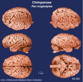
The human brain is by far the most intriguing, complicated, and highly organized organ in the human body. Furthermore, the human brain is far more complex then all other known creatures, stars, galaxies, and planets in the universe. It is no wonder that research on the human brain has been an extremely daunting and challenging task for scientists. However, despite the demanding nature of brain research, scientists have made great progress in understanding the intricacies of the brain. From the teaching of Aristotle to the findings of Broca, advances in brain research have enabled scientists to further understand the functionality of the human brain, and this, in essence, has helped them develop methods of analysis and treatment for illnesses such as schizophrenia, depression, anxiety, and many others.
Historical Findings on the Brain
Throughout history, many have attempted to understand and explain the functionality of the human brain. For instance, in ancient Egypt, the heart and not the brain was regarded as the most important organ within the human body. Furthermore, archeological evidence from 2000 BCE suggests that trepanation, a form of brain surgery that involved cutting a hole through one’s skull, was widely practiced by individuals in prehistoric civilizations. The main purpose of trepanation is not known for certain. However, scientists believe that this practice could have been for religious/mythical rituals or it could have been performed in hopes of relieving one from headaches, epilepsy, and mental illnesses.
In 450 BCE, Alcmaeon, a Greek physician, performed some of the earliest recorded dissections. His work led him to conclude that the brain was the central organ of sensation and thought. In contrast to Alcmaeon’s findings was the great philosopher Aristotle. The latter believed that the heart was the center for sensation, thought, and emotion. At the time, Aristotle’s position on this matter was well respected by many, and his teachings were extremely influential.
 In the early 1800s, German anatomist Franz Gall founded the study of phrenology and became the first person to propose the idea of cerebral localization, a doctrine that emphasized that various mental faculties could be localized to different parts of the human brain. Gall claimed that he could identify and localize 27 faculties in different parts of the human cerebral cortex by simply examining the bumps on one’s skull. As dubious as this practice may seem, it was widely accepted at the time because it offered a method to objectively assess one’s personality characteristics.
In 1848, a horrible accident involving a railroad worker named Phineas Gage enabled researchers to get a better understanding of the concept of cerebral localization and also of the frontal lobe’s role in personality characteristics. Gage was struck with an iron rod, which penetrated his skull and passed through his frontal lobe. Gage survived the incident. However, after recovering, he was no longer the loving and caring person that his friends and relatives remembered. Gage’s personality quickly changed to one that expressed much impulsiveness and anger. The incident led to the suggestion that the frontal lobe of the brain played a significant role in the regulation of emotion.
In 1861, French scientist Paul Broca made a discovery that had a lasting impact on the doctrine of cerebral localization. Through the use of clinical case studies, Broca noticed that patients who suffered from aphasia (a loss or impairment in language) had damage to the left frontal lobe. He concluded that the area responsible for speech is located on the left frontal lobe, on the posterior portion of the third frontal convolution. Although Broca wasn’t the first person to propose that the left frontal lobe was the center for speech, his well-documented clinical studies and his accuracy in pinpointing the exact area (now called “Broca’s area”) is what differentiated his research from those of others such as Gall and Dax.
Nervous System
The human brain weighs approximately 1.3 kg and despite its relatively small size is composed of more than 100 billion neurons. The neurons within the human brain are commonly referred to as the “building blocks” of the nervous system. These neurons consist of dendrites, a cell body (soma), and an axon. Information relating to memory, feeling, impulse, and thought travels from the dendrites to the cell body, then away from the cell body to the axon. Once the information reaches the end of the axon at the site called the “terminal buttons,” the information is released into the synapse by way of synaptic vesicles. These synaptic vesicles carry molecules known as neurotransmitters. Neurotransmitters are found throughout the nervous system and play a key role in carrying information across the synapse. Therefore, neurons communicate by means of several different neurotransmitters, and disruption of these neurotransmitters can have serious adverse effects on the human brain.
The other type of cell found within the human brain is known as the glial cells. These cells outnumber neurons by about 10 to 1. Glial cells are found within the spaces between neurons and provide structure and support for the neuron.
Organization of the Nervous System
The nervous system comprises the central nervous system (CNS) and the peripheral nervous system (PNS). The CNS is composed of the brain and the spinal cord. The spinal cord’s primary function is to carry sensory information to the brain and relay motor instructions from the brain to muscles and glands throughout the human body. The PNS consists of bundles of axons from many neurons that connect the CNS with sense organs, muscles, and glands throughout the body. These “bundles of axons” are commonly referred to as “nerves.” The PNS subdivides into the somatic nervous system and the autonomic nervous system. The somatic nervous system connects the brain and spinal cord to voluntary muscles. The main role of the somatic nervous system is to regulate voluntary muscular movement in response to several external environmental demands. Alternatively, the autonomic nervous system connects one’s internal organs and involuntary muscles to the central nervous system. Furthermore, the autonomic nervous system subdivides into the sympathetic and parasympathetic systems. The sympathetic system prepares the body for rigorous activity; it is also known as the “fight-or-flight” system. This system increases the heart rate, blood pressure, and concentration of blood sugar in the body. On the other hand, the parasympathetic system is known to work in the opposite way. This system enables the conservation and restoration of energy that was dispensed during physical activity. As a mediator, the parasympathetic system decreases heart rate, blood pressure, and concentration of blood sugar. The sympathetic and parasympathetic systems work “hand in hand” to maintain one’s internal bodily functions and homeostasis. The autonomic nervous system is crucial to one’s overall health; without this balancing act, the human body would not be able to properly respond, react, and recover from stressful, strenuous, and frightening stimuli.
Protective Barrier
The human brain is responsible for many essential duties, such as intellect, thought, intuition, memory, emotion, and imagination. Consequently, this organ needs to be well protected from the external environment. Fortunately, the human brain is well sheltered by bone, tissue, and fluid. First, the outermost layer of the human brain is known as the skull; it is a thick, bony structure composed of the occipital, temporal, parietal, and frontal bones. Found within the skull are three protective and supportive tissues known as the meninges. The outermost layer of the meninges is known as the dura matter, a tough and thick membrane that follows the outline of the skull. Next, beneath the dura matter is the thin weblike membrane known as the arachnoid membrane. The third meningeal layer is the pia matter; this tissue is very tightly bound to the surface of the brain and spinal cord. Another essential protective barrier found within the human brain is the cerebral spinal fluid (CSF). This fluid allows the brain to float within the cranium, allowing the brain to withstand certain degrees of physical trauma. Furthermore, it lessens the overall weight of the brain. When floating in the CSF, the brain weighs a mere 50 g instead of 1,400 g. Although the brain does receive trauma through activities such as sports and vehicle accidents, the protective barriers provided by the skull, meninges, and CSF enable the brain to withstand much of the physical trauma. Without these protective tissues, the brain would surely be unable to cope with even minor blows to the head.
Structures Within the Brain
The cerebral cortex is the outer covering of the cerebrum. It is divided into two nearly symmetrical halves, known as the cerebral hemispheres. Although the two hemispheres seem to be two separate and distinctive brains, they are actually attached by a structure known as the corpus callosum. This structure enables the transmission of neuronal messages back and forth between the two cerebral hemispheres. The left hemisphere is in charge of verbal, intellectual, analytical, and thought processes, while the right brain or right hemisphere is in charge of personality, intuition, nonverbal, emotional, and musical functions. Found within the cerebral cortex are the four lobes: frontal, temporal, parietal, and occipital. The brain’s frontal lobe is mainly involved with voluntary motor functions, aggression, and emotion. The right frontal lobe controls the left side of one’s body, while the left frontal lobe controls the right side of one’s body. The temporal lobes are mainly involved with hearing and are conveniently located just above the ears on the sides of each cerebral hemisphere. The parietal lobe is situated at the top of the brain, posterior to the frontal lobe. This area of the brain receives and evaluates certain sensory information that is sent its way. Last, there is the occipital lobe that is located just behind the parietal lobe and is primarily in charge of visual functions.
The brain stem is composed of a group of structures in the brain that regulate bodily functions crucial to the survival of humankind. Within the brain stem is found the medulla oblongata, pons, and midbrain. The medulla oblongata is the lowermost part of the brain stem. It contains several regions that are involved with reflexes, such as swallowing, heartbeat, blood pressure, breathing, coughing, and sneezing. Located just above the medulla oblongata is the pons. The pons is composed of a band of nerve cells about an inch long, which serves as the link between the midbrain and the cerebellum. Furthermore, the pons contains structures that play a role in sleep, arousal, and the regulation of muscle tone and cardiac reflexes. Finally, the upper-most part of the brain stem is known as the midbrain. Located within the midbrain are the superior and inferior colliculi, which are primitive centers concerned with vision and hearing. Furthermore, the midbrain also plays a role in the perception of pain and the guidance of movement by sensory input from vision, hearing, and touch.
The thalamus is situated just above the brain stem very close to the center of the brain. The yo-yo shaped thalamus receives input from all senses with the exception of olfaction and then directs these messages to the cerebral cortex and other parts of the human brain.
The hypothalamus is a tiny structure that plays a major role in influencing behavior primarily through the production and release of hormones. The hypothalamus is involved with the regulation of the autonomic nervous system by helping control heart rate, movement of food through the digestive tract, urine release from the bladder, perspiration, salivation, and blood pressure. Furthermore, it also contains centers that influence consumption of food and water, reproduction, metabolism, homeostasis, and the regulation of the sleep-wake cycle. Last, the hypothalamus is connected to the pituitary gland and plays a role in the regulation of the endocrine system.
The cerebellum is situated just behind the brain stem and weighs approximately one eighth of the brain’s total mass. It is primarily involved with the coordination of muscular activities. It has also been found to play an important role in emotions such as anger and pleasure.
The limbic system is composed of several structures that play a role in emotional responses and behavior. One of the structures found deep within the human brain is the amygdala. This part of the limbic system is important for fear, anxiety, and aggression responses. Another important part of the limbic system is the hippocampus; this structure is linked to the hypothalamus and is very important for memory functions.
The basal ganglia is located on the left and right sides of the hypothalamus. It is composed of the caudate nucleus, putamen, and globus pallidus. These structures work together to exchange information with the cerebral cortex. Its main role is to plan motor sequences of behavior.
Technological Revolution
Throughout the years, much progress has been made in understanding the structure and function of the human brain. Most of the progress in understanding the complexities of the human brain derive from the advances in neuroimaging techniques. Neuroimaging can be divided into two basic categories, structural imaging and functional imaging. The former involves a scanning technique that reveals the gross anatomy of the human brain. Examples of structural neuroimaging include computerized tomography (CT) and standard magnetic resonance imaging (MRI). On the other hand, functional imaging is used to provide information on some sort of functional brain activity. Such imaging techniques are provided by positron emission tomography (PET), single photon emission tomography (SPECT), and functional magnetic resonance imaging (/MRI). Researchers use these instruments to measure certain aspects of brain activity, such as cerebral blood flow and oxygen consumption. With the help of functional and structural imaging techniques, surgeons in the field of neurology can fully examine the brain prior to performing dangerous surgical procedures.
Mental Disorders and Imaging Techniques
In the past, mental disorders were very difficult to classify, and many believed that mental disorders were nothing but a human weakness. Furthermore, many believed that those who suffered from a mental illness were possessed by evil spirits and thus were placed in hospitals for the mentally insane, where they were often deprived of their basic human rights. However, many of these beliefs have changed since the arrival of technological instruments that could measure the structural and functional anatomy of the human brain. Such techniques have illustrated that mental diseases such as schizophrenia, autism, and attention-deficit hyperactivity disorder may in fact have biological aspects.
For instance, researchers have found that decreased frontal lobe volume and diminished neuronal and blood flow activity are common in patients who suffer from schizophrenia. Furthermore, MRI studies suggest abnormalities in the temporal lobes of individuals afflicted with schizophrenia.
There has also been an increasing amount of research on autism. Researchers have found that defects in the brain stem may account for symptoms of the disorder and also that the cerebellum and frontal lobes are less developed in autistic patients than in normal individuals.
Another popular and often misunderstood mental disorder is attention-deficit hyperactivity disorder or ADHD. Researchers have been able to identify structural abnormalities among ADHD patients. For instance, it has been found that ADHD patients seem to have smaller corpus callosums and caudate nucleii than those patients without the disorder.
Therefore, neuroimaging techniques have enabled researchers to further understand mental disorders, and the knowledge gained from these techniques can help in developing methods of treating, rehabilitating, and caring for those affected by these mental disorders.
The human brain is a highly complex and intriguing organ that has proven to be very difficult to study. However, with the pioneering research of many individuals within the wide range of disciplines, such as chemistry, biology, psychology, medicine, and others, much knowledge has been gained about the functional and structural characteristics of the human brain. Although researchers have discovered many of the mysteries of the human brain, there remains much to be discovered. Through a multidisciplinary approach, advances in medical technology, and more government funding toward research, remaining secrets of the human brain will begin to be unraveled.
References:
- Beatty, J. (1995). Principles of behavioral neuroscience. Dubuque, IA: Wm. C. Brown.
- Joel, D. (1997). Mapping the mind. Toronto, Canada: Carol Publishing.
- Nietzel, M., Bernstein, D., & McCauley, E. (1997). Abnormal psychology. Englewood Cliffs, NJ: Prentice Hall.
- Purves, D., Augustine, G., Fitzpatrick, D., Katz, L.,LaMantia, A-S, McNamara, J., & William, S. M. (2001). Neuroscience (2nd ed.). Sunderland, MA: Sinauer.
- Restak, R. (2001). The secret life of the brain. Washington, DC: Joseph Henry Press.
- Springer, S., & Deutsch, G. (1997). Left brain rightbrain (5th ed.). New York: W.H. Freeman.
- Toomela, A. (Ed.). (2003). Cultural guidance in the development ofthe human mind. Westport, CT: Ablex.

